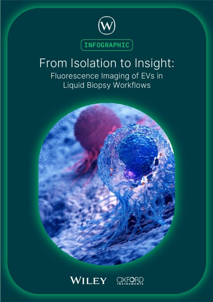
From Isolation to Insight: Fluorescence Imaging of EVs in Liquid Biopsy Workflows
This infographic hopes to provide scientists with a comprehensive overview of fluorescence imaging strategies for extracellular vesicle research, allowing you to select the optimal combination of labeling techniques, imaging modalities, and analysis workflows to advance your liquid biopsy and cancer diagnostics research.
Register now to receive this complimentary infographic!
What you will learn about:
- Understand the clinical imperative driving EV research in liquid biopsy applications
- Compare traditional EV characterization methods with fluorescence imaging approaches
- Select appropriate fluorescent labeling strategies for EVs (lipophilic dyes, immunofluorescence, covalent labels)
- Match imaging modality to research question: widefield for screening, confocal for 3D context, super-resolution for nanoscale features
- Integrate multi-scale imaging into a six-step EV workflow from isolation to analysis
- Leverage automated workflows for EV detection, counting, and tracking in complex datasets
- Recognize key technical requirements for reproducible EV imaging (laser lines, detection sensitivity, quality control)
More Information
Extracellular vesicles (EVs) are transforming liquid biopsy diagnostics, offering unprecedented insights into cancer detection and monitoring. However, imaging EVs at the nanoscale while distinguishing them from artifacts remains a significant challenge. This infographic guides researchers through fluorescence microscopy strategies for EV characterization—from labeling approaches to multi-modal imaging techniques and automated analysis workflows. Discover how to integrate widefield, confocal, and super-resolution imaging into your EV research pipeline to achieve rapid detection, 3D tissue context, and nanoscale resolution of EV subpopulations.
Access
Visit our Content Hub, register your details to create a user profile for the hub, then login to access all content within the hub.
Content Hub Access